


Digital x-rays
 Using digital X-rays imaging allows for a multitude of advantages. These advantages include the ability to transfer and enhance images. Due to the digital components of these machines we are able to bypass the chemical processing of the traditional photographic film, and the time it takes to process is greatly reduced.
Digital radiography uses a digital image capture device, which allows for immediate availability of the image; as well as the ability to use special techniques to enhance the overall image to insure better image quality.
As with any bone related problem, a dental X-ray is necessary when any form of pain exists in the mouth that requires treatment. The source of pain can be either an infected tooth or a problem in the periodontal tissues surrounding the teeth, i.e. bone and gums. In any case, to reach an accurate diagnosis, an X-ray will be required to assess the source of infection and pain in order to treat the dental problem accordingly.
In current dentistry, X-rays are taken digitally rather than on film, so that the X-ray image of the tooth and bone is produced immediately for examination. It is faster, more detailed and can also be manipulated on a computer screen for better visualization. Conventional X-rays also tend to fade out on film after a period of time, which does not apply to digitally taken X-rays which can be saved and stored on a computer database easily. Furthermore, X-rays taken conventionally also require a higher amount of radiation emitted, which a digitally taken X-ray produces far lesser of.
Digital X-rays can be taken intra-orally (within the mouth) or extra-orally (outside the mouth). Intra-oral X-rays are taken in different positions, such as the following:
- Periapical X-rays: allows the whole tooth to show in an X-ray image along with the surrounding bone. Shows the presence of any lesion on or around the roots of teeth.
- Bitewing X-rays: used primarily to check for presence of a lesion between teeth, be it a cavity between the teeth or the loss of bone between the teeth in periodontal disease.
- Occlusal X-rays: show all teeth on a particular side from the chewing surfaces.
Extra-oral X-rays are used to assess the bony structures in the skull along with the jaws, useful for when a bigger picture of the facial skeleton is needed rather than a few teeth:
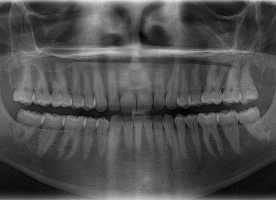
- Panoramic X-rays or OPG: shows both jaws with all teeth (including un-erupted teeth present within the jaw bones), along with the temporo- mandibular or jaw joint (TMJ).
- Cephalogram: shows the lateral view of the entire facial skeleton including the first few vertebrae, very useful in orthodontic treatment.
- CT scans: shows 3D images of the bones, useful for assessing the presence of fractures or tumors within the bones.
- Sialograms: To assess any abnormality in the salivary glands.